Uni Wien:Neurolinguistik VO (Reiterer)/Fragenkatalog SS18
Approximately 60+~ factual questions (no essay style necessary) about the most important basic concepts, scholars, inventors, techniques, disorders.... (concepts again detailed below), plus knowing what some important acronyms stand for, plus being able to graphically position some basic neuroanatomical landmarks in a schematic brain. Since the field of cognitive neuroscience nowadays uses a lot of acronyms, it is important to know some of them, so please look at the acronyms again. All in all, a good overview and not knowing every detail should be achieved.
The concepts according to their chronological occurrence during the lecture: You should be able to name always some important representatives of the following “concepts” or issues: aspire to remember 5-10, at least 3 of every kind.
Die 15 Fragen des Katalogs sind im Folgenden als Abschnittsüberschriften aufgeführt - bitte weiter ausfüllen und ausarbeiten:
Journals of Cognitive Neuroscience, especially Neurolinguistics[Bearbeiten | Quelltext bearbeiten]
to be studied for the exam: "You should be able to name some of them, of course not all, maybe some 5."
| typical journals in Neurolinguistics | typical journals in Cognitive Neuroscience | Impact Factor |
|---|---|---|
| Brain and Language (neurobiol. of lang.conference, NSL) | Nature | 38 |
| NeuroImage | Nature Neuroscience | 15 |
| Human Brain Mapping | Nature Reviews Neuroscience | 32 |
| Journal of Neurolinguistics | Science | 32 |
| International Journal of Language and Communication Disorders | Trends in Cognitive Sciences | 17 |
| Language, Cognition and Neuroscience | Trends in Neurosciences | 14 |
| Aphasiology | Neuron | 16 |
| Bilingualism: Language and Cognition | Cortex | 6 |
| Mind, Brain and Education | Cerebral Cortex | 7 |
| Frontiers in Language Sciences /Psychology | Cognition | 3 |
| Psychological Bulletin | 16 | |
| PloS Biology | 13 | |
| PNAS | 10 | |
| Current Opinion in Neurobiology | 7 | |
| Journal of Neuroscience | 7 | |
| Journal of Cognitive Neuroscience | 4 | |
| Brain Research | 3 | |
| Cognitive Brain Research | 3 |
Scimago journal ranking in language & linguistics
The "neutral" database: PubMed
Some search engines for looking up publications in the field:
Some important organizations with annual conferences:
Basic tissue substances in the brain[Bearbeiten | Quelltext bearbeiten]
outer to inner:
- Cerebro-Spinal Fluid (in ventricle system)
- Gray Matter (cell bodies in the cortex)
- White Matter (myelinated nerve fibers)
Cytoarchitecture[Bearbeiten | Quelltext bearbeiten]
to be studied for the exam: "E.g. Golgi, Brodmann, ..some important historic figures and what they achieved."
Cytoarchitecture (="cell architecture") refers to the cellular composition of brain tissue (see also histology)
Cell Types[Bearbeiten | Quelltext bearbeiten]
to be studied for the exam: "Should be able to name some cell types"
- Neurons: transmit messages (via dendrites, synapses, axons → electro-chemical signals); ca. 86 billion, each connected to up to 10,000 other neurons through 100-200 k synaptic connections per neuron, totaling up to 1 trillion).
- by form
- unipolar: only 1 "process" [~appendix]
- bipolar: 2 processes: 1 axon, 1 dendrite
- multipolar: 1 axon, 2+ dendrites
- by function
- afferent (sensory neurons): from organs to CNS/brain (central nervous system = ~brain + spinal chord)
- efferent (motor neurons): from CNS to peripheral nervous system → effectors (cells that actively respond to a stimulus and effect some change, e.g. muscles, glands but also microglia in brain and spinal chord)
- interneurons: connect within CNS
- by form
- Glia ("glue"): supporting functions [think housekeepers]: maintain homeostasis (~steady state of physico-chemical conditions), form myelin, fixate, insulate, and protect neurons, support them with nutrients and oxygen, destroy pathogens and remove dead neurons
- Macroglia
- Astrocytes (astroglia): Regulate neurotransmitter concentrations; important for blood-brain barrier
- Oligodendrocytes: (oligo = few, dendron = tree) enwrap axons → make up myelin sheaths
- Microcytes (microglia): macrophages (type of white blood cell) which remove plaques, dead neurons, all types of foreign bodies [clean up and defend against threats]
- Macroglia
- other cell types
- Pyramidal cells:
- basket cells
- Betz cells (large motor neurons)
- Purkinje cells (large cells in cerebellum)
Neuroanatomy[Bearbeiten | Quelltext bearbeiten]
Presentation of a "fresh dead brain"
Brain
- ~ 1.5 kg
- 86 billion neurons
- each connected to up to 10,000 others
- over 1,000 trillion (1 quadrillion) connections via synapses (up to a quadrillion according to the lecture slides, apparently quoted from this scantily sourced site)
- accounts for 20 % of total glucose consumption (~400 kcal/day)
- 20 % of total oxygen consumption
Systematics/taxonomy by:
- morphological structure (shape) (e.g. amygdala)
- evolutionary age (e.g. Neocortex, Archicortex, brain stem ("reptilian brain")
- location (e.g. frontal lobe)
- cell structure (cytoarchitecture, physiological function)
- physiological & sensory function (e.g. primary audio cortex)
- higher order (cognitive) function (e.g. peri-sylvian language network, default mode network, limbic system ("emotional circuit"), reward system ("pleasure loop)
- name (e.g. Broca's area, sylvian fissure, Rolandic fissure)
Basic global structure[Bearbeiten | Quelltext bearbeiten]
to be studied for the exam: "Parts of the brain and their most basic functions. At least one function per global brain part (e.g. thalamus, limbic system...)."
4 directions:
- dorsal (superior): top side (towards the back [cp. quadripedal animals, where the back is on top/facing away from the ground; also, dorsal referring to the top side of the tongue (e.g. dorsal fricatives)])
- ventral (inferior): bottom side ("bellywards" [cp. quadrupeds, abdomen is on underside])
- rostral (frontal, anterior): front side ("beakwards")
- caudal (posterior): back side ("tailwards")
Surrounding layers: Dura Mater (outer + inner), Arachnoid, subarachnoid space, Pia Mater
4 important parts: cerebrum, cerebellum, diencephalon, brain stem
- Cerebrum (Großhirn, Telencephalon): left & right hemisphere;
- Corpus callosum: Main fiber connection between hemispheres; largest white matter structure in the human brain, consisting of 200-300 million axonal projections
- Cortex (Hirnrinde → outer layer of Cerebrum, consiting of gray matter) vs. subcortical areas (embedded within white matter → myelin-sheathed; composed of axons (nerve fibers))
- Frontal Lobe: reasoning, motor skills, higher level cognition, and expressive language
- Prefrontal Lobe
- Parietal Lobe: tactile sensory information such as
- Superior Parietal Lobe
- Inferior Parietal Lobe
- Occipital Lobe: primary visual cortex
- Temporal Lobe: primary auditory cortex
- Hippocampus: memories, learning
- Cerebellum (Kleinhirn): 10 % of brain volume, 50 % of neurons
- important for
- motor control & coordination (gait & posture)
- fine motor control (balance, motor learning)
- learning, cognition, speech (tempo, pace [speech motor functions, rhythm])
- structure
- mid portion
- Vermis cerebelli ("worm"):
- cerebellar hemispheres
- contains
- arbor vitae:
- folia:
- Purkinje cells
- important for
- Diencephalon (Zwischenhirn)
- Thalamus: Very important regulative system! A bilateral complex of cell nuclei, which have fiber connections to the cerebrum. The Thalamus acts as a switchover point for most sensory pathways → every sensory system (except olfactory) has a its own thalamic nucleus, which relays sensori-motor signals to the cortex. The Thalamus regulates consciousness, sleep and alertness
- Epithalamus
- Subthalamus (globus pallidus (Pallidum), capsula interna)
- Hypothalamus: regulates vegetative functions: blood pressure, temperature, circadian rhythm, sexuality, eating, drinking
- Brain stem (Hirnstamm)
- Medulla oblongata ("verlängertes Rückenmark") → ARAS (ascending reticulary formation): basic functions
- sleep/wake system (through emitting serotonine, adrenaline, noradrenaline)
- breathing, larynx, swallowing, coughing, vomiting [autonomous (involuntary) functions)]
- Pons relay station for motor and sensory innvervation and connections between cerebellum & cerebrum (Kleinhirn & Großhirn)
- Mesencephalon (mid brain): transition unit for sensory info from eye and ear to brain
- substantia nigra: important role in reward system (produces neurotransmitters, esp. dopamine) and movement; Parkinson's disease
- PAG (peri-aqueductal gray): part of endorphine system
- Medulla oblongata ("verlängertes Rückenmark") → ARAS (ascending reticulary formation): basic functions
- Limbic system: Formation of memories, emotional life
- Amygdalae: 2 almond shaped clusters within the temporal lobes, primary role in memory processing, decision-making, emotional responses (fear, anxiety, aggression)
Gyri & Sulci (fissures)[Bearbeiten | Quelltext bearbeiten]
to be studied for the exam: "Especially those related to language processing"
Sulci
- Sulcus Centralis = Fissura Rolandi (rolandic fissure) (abbrev.: cs)
- Sulcus lateralis = Fissura Sylvii (sylvian fissure)
- ifs: Inferior Frontal Sulcus
- ips: Intraparietal Sulcus
- sts: Superior Temporal Sulcus
Gyri
- Heschl's Gyrus (BA 41, 42)
- located in Primary & Secondary Auditory cortex at Sylvian Fissure, in front of (and seperate from) the Planum Temporale
- Planum Temporale (BA 22)
- Superior Temporal Gyrus (STG) (BA 22, 41, 42)
- Inferior Frontal Gyrus (IFG)
- Angular Gyrus (BA 39)
- Supramarginal Gyrus (BA 40)
- Inferior Frontal Grus (BA 44, 45)
- Inferior Parietal Lobe (IPL)
Brodmann Areas[Bearbeiten | Quelltext bearbeiten]
to be studied for the exam: "The most important ones, here some 10."
3 parcellation approaches:
- Functional: Cyto-architecture and myelo-architectonic structure
- Receptor-architectonic structure (receptor binding, density of neurotransmitters)
- Connectivity-based parcellation approach
| BA # | Name | Notes | short description (fill in from slides & other sources) | |
|---|---|---|---|---|
| important areas
(speech/language) |
39 | angular gyrus | IPL | |
| 40 | supramarginal gyrus | IPL | ||
| 41 | Heschl's gyrus | longer and wider in LH → more underlying myelin, greater interconnectivity → faster transmission) | Primary Auditory Cortex | |
| 42/22 | planum temporale/secondary auditory areas | |||
| 22, 21, 42 | superior temporal gyrus | |||
| 22 (in dominant hemisphere!) | Wernicke's area | perception of speech: hearing, listening, receiving, understanding; sensoric aspects | ||
| BROCA | (in dominant hemisphere!) | Syntax | production of speech: speaking, articulation; motoric aspects | |
| 44, 45, 47 | inferior frontal gyrus | |||
| 44 | pars opercularis | |||
| 45 | pars triangularis | |||
| 47 | pars orbitalis | |||
| important areas
(general) |
04 | (primary) motor cortex | ||
| 01, 02, 03 | somatosensory cortex | |||
| 6 | premotor cortex + SMA (supplementary motor area) | neurons of SMA project directly to the spinal cord | ||
| 17 | primary visual area | Cuneus | ||
| 18 | secondary visual area | |||
| 19 | tertiary visual area | |||
| 46 | dorsolateral prefrontal cortex | |||
| 23 | cortex cingularis posterior ("cingulum") | |||
| 21 | middle temporal area of cortex | |||
| 07 | superior parietal lobule+precuneus | |||
| 10 | anterior/fronto-polar prefrontal cortex | |||
| 08 | frontal eye fields | |||
| 09 | frontal cortex | |||
| 13 | insular cortex | interoceptive awareness, expressive (Broca's) aphasia |
Primary sensory systems[Bearbeiten | Quelltext bearbeiten]
to be studied for the exam: "Vision, audition, motor control (homunculus), “-topy”-principles (e.g. tonotopy)"
Vision[Bearbeiten | Quelltext bearbeiten]
PLEASE FILL IN
Audition[Bearbeiten | Quelltext bearbeiten]
PLEASE FILL IN
Motor Control[Bearbeiten | Quelltext bearbeiten]
PLEASE FILL IN
"-topy"-principles[Bearbeiten | Quelltext bearbeiten]
Topologically represented, contiguous processing areas in the brain
- Tonotopy: Frequency-specific sound-processing areas → Tones close to each other in terms of frequency are represented in topologically neighbouring regions in the brain (esp. Heschl's gyrus).
- Retinotopy: Retinal mapping, i.e. mapping from retinal areas to brain areas. The mapping is not strictly contiguous, i.e. upper half of retina is processed seperate in a region seperate from the one processing the lower half of the retina/visual field
- Somatotopy: Point-for-point correspondence of an area of the body to a specific point of the brain, esp. somatosensory complex
- Syntactotopy? To what extent can syntactic processing be mapped to specific brain areas?
Differences between the hemispheres (lateralization)[Bearbeiten | Quelltext bearbeiten]
to be studied for the exam: "Structural[] and functional[] [differences between hemispheres]."
Visual and somatosensory information is predominantly processed in the hemisphere opposite the originating side of the body and visual field.
Bouillaud 1825, Gall 1835, M. Dax 1836
Broca 1861: Speech (and its loss: "aphemia") is connected to the third convolution of the superior part of the left frontal lobe
Hughlings Jackson[Bearbeiten | Quelltext bearbeiten]
1870: …found that the inactivation of the right hemisphere (RH) causes a monotonous, affectless character of the patient‘s speech together with the inability to regulate the voice according to the degree of the situational emotivity.
In this situation, where the left hemisphere “talks alone”, the patient shows greater talkativeness, because the language act is not controlled or regulated by the right hemisphere. …Thus such a patient understands the “denotational meaning of what is said, but fails to recognize, whether it is spoken in an angry, sad or humorous way…
…In contrast, a patient with a damaged Left hemisphere (LH) will neither comprehend what is said, nor be able to produce coherent, intelligible speech, but is still able to comprehend and produce the emotional tone…
If LH is lesioned or destroyed anteriorly, patient retains emotional language
..“over one hundred years ago scientific intuition enabled the sagacious J H Jackson to assign the intellectual language to the left and the emotional language to the right hemisphere“…
(Roman Jakobson)
---
- contra-laterality (antonym: ipsilaterality): visual and auditory stimulus processing as well as [volitional] motor control of many parts on the right side happens in left hemisphere and vice versa, e.g. right arm & leg, right half of visual field (of both eyes!), right ear in left hemisphere
- optical nerves are split between left and right half of the retina; nerves carrying signals from the inner half of each retina (i.e. left half of right eye's retina and vice versa) cross over at the Chiasma opticum, which results in signals from the left half of both retinas being routed to the left hemisphere and vice versa - nervertheless, the left half of the visual field is processed in the right hemisphere since the "light rays" also cross over due to refraction in the cornea (lens of the eye)
- left and right motor impulse pathways cross over in the Medulla oblongata
- sidedness
| left hemisphere | right hemisphere |
|---|---|
| speech, language, comprehension | creativity |
| analysis and calculations | spatial orientation |
| time and sequencing | context/perception |
| recognition of words, letters and numbers | recognition of faces, places and objects |
| memory? | music |
| emotion? | |
| visual perception?? | |
| initiation of movement | maintenance of ongoing movement |
| intellectual language | emotional language |
| language in general | visuo-spatial processing
emotional processing |
| digital, serial, sequential | analogue, parallel, synchonous |
| linear | complex |
| details | global picture |
| analytic | holistic |
| rational | intuitive |
| abstract | concrete |
| conscious, active | unconscious, automatic |
| fine motor coordination | gait, posture |
| analytical processing of music | emotional processing of music |
| time | melody, sound, melody, pitch (better pitch resolution necessary for music than for language (~1/2 octave for language, 1/6-1/12 octave for music) |
| short sounds (20-50 ms) <- higher time resolution necessary for language compared to music) | long sounds (>50 ms) |
| line drawings | pictures, faces |
| linear, abstract schemes | complex, spatial figures |
| syntax, morphology, semantics | pragmatics, discourse, coherent text |
| abstract words | concrete words |
| function words, verbs | content words, simple nouns & adjectives |
| propositional language, new sentences | ritualized formulae, chunks of words |
| denotation | connotation, metaphor |
| linguistic meaning | social context of linguistic communication |
| phonetics & phonology | foreign accent detection |
| minimal pair distinction, syllables | sentence intonation, melody of speech, prosody |
| linguistic prosody | affective prosody |
| speaking | singing |
| alphabetical script | ideographic [e.g. Chinese], pictographic script (e.g. traffic signs) |
PET study: processing spoken words in different accents (“bale”, “dale”, “pail”, tail” in Australian, British, French, Hindi)
Results: Detection of words -increased blood flow in a left frontal area associated with motor and phonetic processing, and a left temporal area associated with semantic memory.
Homologous areas of the right hemisphere, together with right prefrontal and precuneus regions, showed increased blood flow during detection of accents.
Interpretation: LH specialization for extraction of the linguistic, phonetic and semantic information contained in speech,
and RH specialization for pragmatics, the social context of linguistic communication, prosody, accents.
→ Both hemispheres needed in normal communicative situations
Vallortigara & Rogers (2005): Survival with an asymmetrical brain: advantages and disadvantages of cerebral lateralization
| Vallortigara & Rogers (2005): Survival with an asymmetrical brain: advantages and disadvantages of cerebral lateralization | |
|---|---|
| Left Hemisphere | Right Hemisphere |
| prey discrimination and catching (fish, toads) | predator detection (fish, chicks) |
| foraging with discrimination and/or manipulation of food items (birds) | predator escape (frog tadpoles, fish, toads, chicks, dunnarts) |
| approach and manipulation of objects (birds, monkeys, apes) | neurochemical changes with predator stress (rats, cats) |
| avoidance/withdrawal (monkeys, apes, humans) | |
| inhibition of aggression (chicks, humans) | fear (chicks, rats) |
| inhibition of intense emotions, especially negative emotions (humans) | aggression (toads, lizards, chicks, monkeys) |
| courtship and copulatory behavior (newts, birds) | |
| expression of intense emotions (monkeys, apes, humans) | |
| recognition of categories/attention to large changes (birds, rats) | contact/monitoring of conspecifics (fish, tadpoles) |
| recognition of species-typical vocalizations (birds, mice, some monkeys, humans for speech) | recognition of individual conspecifics (chicks) |
| attention to landmarks (birds) | spatial cognition (birds, rats, humans) |
| attention to locals cues (birds, monkeys, humans) | attention to global cues (chicks, monkeys, humans) |
| Considered responses -> able to inhibit responding while deciding between alternative responses | Rapid, species,typical responses |
| visuo-spatial analysis centered on local features | visuo-spatial analysis centered on relational properties of the spatial layout |
- handedness/dominance: In left-handed people some functions are mostly located in the other hemisphere (as compared to the list above), especially relating to language
- interconnection: Fast Interaction by commissure fibers (connection fibers between the hemispheres)
- corpus callosum (CC)
- smaller CC associated with schizophrenia and Tourette's (both related to verbalization and language)
- bigger CC associated with ambidexterity (ambidextrous people have better memory and can recall events from younger ages)
- commissura anterior (AC)
- subcortical fibers
- corpus callosum (CC)
- redundancy: Each hemisphere is a relatively complete "brain" to itself, with a broad range of abilities
Experiments & studies[Bearbeiten | Quelltext bearbeiten]
Roger Sperry[Bearbeiten | Quelltext bearbeiten]
Split-brain patients (CC surgically severed to treat epilepsy) can only repeat words shown in right half of visual field because they are processed in the left hemisphere (which also covers the right half of the visual field - contra-laterality!) and the connection between hemispheres is cut; words shown in the left half of the visual field can be drawn, however.
WADA test[Bearbeiten | Quelltext bearbeiten]
Processing in one hemisphere is inhibited by injecting a barbiturate into the corresponding carotid arteria. If the predominantly language-processing hemisphere is inhibited (LH in 96 % of right-handed subjects and in 70 % of left-handed subjects, RH in 4 % of right-handed and 15 % of left-handed), objects given to the test subject cannot be named but indicated on a chart.
Knecht et al.[Bearbeiten | Quelltext bearbeiten]
Comparison of neuronal activation in relevant areas of LH vs. RH during word generation (by measuring blood perfusion with Doppler sonography).
Result: Lateralization was equivalent in men and women. (iii) Right hemisphere dominance was found in 7.5% of subjects. These findings indicate that atypical language dominance in healthy right-handed subjects of either sex is considerably more common than previously suspected.
An example[Bearbeiten | Quelltext bearbeiten]
| J | J | ||
| J | J | ||
| J | J | J | J |
| J | J | ||
| J | J |
A large letter H (the global level) made up of small letter J ’ s (the local level). When patients with left-hemisphere damage try to reproduce such figures, the intact right hemisphere correctly produces the global pattern (the large H), but ignores the small local details (the small J ’ s). In contrast, in patients with right-hemisphere damage, the intact left hemisphere correctly produces the local detail (several small J ’ s) but does not arrange them into the correct global pattern.
In neurologically intact individuals, global and local levels are processed better when flashed briefly to the left visual field (right hemisphere) and right visual field (left hemisphere), respectively, and functional neuroimaging studies show that attending to global versus local levels produce greater activation in right and left hemispheres, respectively.
(from Hellige (2008))
Auditory cortex - L/R differences[Bearbeiten | Quelltext bearbeiten]
Anatomical differences[Bearbeiten | Quelltext bearbeiten]
- Sylvian fissure longer in LH
- Planum temporale bigger in LH
- Heschl's Gyrus (primary auditory) differs between hemispheres
- Inferior frontal gyrus (Broca) bigger & more indented [in LH?]
- Inferior parietal lobe more convoluted [in LH?]
Asymmetric Sampling in Time (AST)[Bearbeiten | Quelltext bearbeiten]
The ‘asymmetric sampling in time’ (AST) hypothesis developed here provides a framework for understanding a range of psychophysical and neuropsychological data on speech perception in the context of a revised cortical functional anatomic model.
The AST model is motivated by observations from psychophysics and cognitive neuroscience that speak to the fractionation of auditory processing, in general, and speech perception, in particular. Building on the observations:
- that the speech signal contains more than one time scale relevant to auditory cognition (e.g. time scales commensurate with processing formant transitions versus scales commensurate with syllabicity and intonation contours),
- and that speech perception is mediated by both left and right auditory cortices, AST suggests a time-based perspective that maintains anatomic symmetry while permitting functional asymmetry.
AST proposes that the input speech signal has a neural representation that is bilaterally symmetric at an early representational level.
Beyond the initial representation, however, the signal is elaborated asymmetrically in the time domain: left auditory areas preferentially extract information from short (∼20–40 ms) temporal integration windows. The right hemisphere homologues preferentially extract information from long (∼150–250 ms) integration windows. It is suggested that temporal integration is reflected as oscillatory neuronal activity in different frequency bands (gamma (LH, >50 Hz, fast), theta (RH, 4-7 Hz, slow)).
Acoustic uncertainty principle[Bearbeiten | Quelltext bearbeiten]
[Think of measuring your pulse by counting your heartbeats over a span of time. If you count for a whole minute, you get your exact pulse in bpm (beats per minute). If you only count your heartbeats for a few seconds, you can only roughly estimate your bpm but you can make several of these estimates over the span of a minute, which, for example, allows you to notice how your pulse drops whithin the first minute of resting after your work-out.
The longer each measurement period, the more accurate (~minimized average deviation) the average frequency of the signal calculated from each measurement period. In an acoustic signal, this means better pitch detection. On the other hand: Longer measurement periods mean fewer measurement periods per unit of time, which entails lower temporal resolution: Frequency/pitch changes within a single measurement period are not detectable, only changes from one measurement to the next are. Shortening the duration of measurement periods entails less accurate pitch detection from each measurement period but since there are more measurement periods per unit time, temporal resolution increases and faster pitch changes become detectable].
The trade-off between pitch detection accuracy vs. temporal resolution might (at least in part) explain why and how work-sharing and different specializations of the hemispheres is advantageous.
Why lateralization?[Bearbeiten | Quelltext bearbeiten]
- work sharing is more efficient than redundant processing of the exact same things in both hemispheres [robustness by redundancy is still given to a certain degree through autonomy and plasticity of hemispheres]
- -> specialization on different aspects of the same domain, e.g. AST -> fast transients in LH, accurate pitch in RH
- interhemispheric transport time is costly in big brains (to the extent that symmetric processing isn't justified)
- singular organs: only one mouth/vocal tract -> split of control of single organs between hemispheres might be disadvantageous (in contrast to pairwise parts like limbs)
- perfectly symmetric evolution of pairwise organs/parts is highly unlikely -> over time, asymmetries and specializations arise [esp. in internal organs, which do not directly interact with the environment]
Subcortical structures[Bearbeiten | Quelltext bearbeiten]
to be studied for the exam: "Know and name some subcortical structures"
- Thalamus
- Tectum
- Superior & Inferior Colliculus
- Capsula Interna
- Claustrum
- Basal Nuclei/Ganglia (subkortikale Kerne): A group of large nuclei that partially surround the thalamus, important for motor control
- Nucleaus Caudatus (speech)
- + Putamen = Striatum
- Putamen (speech)
- Globus Pallidus (Pallidum)
- Nucleaus Caudatus (speech)
- Nucleus accumbens
- Substantia Nigra
- Nucleus Subthalamicus
- Amygdala ("Mandelkern"), part of limbic system
- motor movement
- Cingulate Cortex
- Corpus Callosum
- Fornix
- Olfactory Bulb
- Mammillary body
- Massa Intermedia
- Fornix
Limbic System[Bearbeiten | Quelltext bearbeiten]
supports a variety of functions incl. motor, behavior, motivation, long-term memory, olfaction; most of emotional life and important for formation of memories
produces endorphines
- Hippocampus
- Gyrus Cinguli
- Basal Ganglia
- Amygdalae: motor movement (basal ganglia)
- Nucleus Accumbens
- Thalamus
- Hypothalamic nuclei
- Fornix
Fiber tracts (fasciculi, white matter fibers)[Bearbeiten | Quelltext bearbeiten]
to be studied for the exam: "Know some fiber tracts (fasciculi) important for language and some general ones."
- Fasciculus Arcuatus
- 3 segments: long, anterior, posterior
Historical figures in neurolinguistics[Bearbeiten | Quelltext bearbeiten]
to be studied for the exam: "E.g. Wernicke, Dax, Lichtheim, Gall... You should also relate their biggest discoveries / theories or models."
- Carl Wernicke (1848-1905)
- 68 pp. monograph "The Aphasic Symptom Complex" in 1874
- wanted to anchor the functional architecture for speech in neural architecture
- wrote "Lehrbuch der Gehirnkrankheiten" at Charitè Berlin
- believed psychiatric illnesses can be based on neurological problems → psychoses can be reversible
- theory of projection, topography of sensory and motor pathways into and out of the brain
- Pierre Paul Broca (1824-1880), son-in-law of Bouillaud (→ 500 francs bet)
- introduced term "limbic system"
- concluded from cases with lesion of left frontal lobe and SLI that language is localized in the left hemisphere
- article published in the same year (1865) as Gustave Dax' article with the same conclusion → Dax accused him of plagiarism
- popularized localism (vs. equipotentialism), which led to research into lateralization
- Broca's area was named by Ogle in 1867
- Sigmund Exner (1848-1926)
- Austrian physiologist, head of med. department of physiology at Uni of Vienna
- cohort theory of spoken word perception (almost a century earlier than others)
- considered neuronal networks as learning and processing mechanisms
- Korbinian Brodmann (1858-1918)
- dt. Neuroanatom und Psychiater
- von Alois Alzheimer zur Neurologie ermutigt
- famous for distinguishing 52 regions of the cortex based on their cytoarchitecture, the Brodmann areas
- published early brain atlases (based on his eponymous areas)
- worked on and findings about insomnia, sleep centers & regulation; intelligence, expertise & exceptional brains
- Camillo Golgi (Nobel prize in medicine, 1906 with y Cajal)
- developed silver staining technique for visualizing nervous tissue in light microscopy ("Golgi's method")
- Santiago Ramón y Cajal
- discovered axonal growth cone and demonstrated that neural network is contiguous, not continuous
- Marc Dax (1770-1837) & his son Gustave Dax (1815-1874), French physicians
- Marc Dax accumulated statistics about 40+ hemiplegic patients (paralysis) with loss of word memory
- loss of speech only in patients with right hemiplegia (left hemisphere!)
- Gustave Dax: 370 cases of hemiplegia
- assumed medial left lobe must be "the seat of articulated speech" but his paper was rejected by the French Academy of Science because they regarded it as mere phrenology, hence pseudoscience
- Marc Dax accumulated statistics about 40+ hemiplegic patients (paralysis) with loss of word memory
- Lichtheim
- Adolf Kussmaul (1822-1902)
- Jean-Baptiste Bouillaud (1796-1881) established the French phrenological society ("phrenology without bumpy organs or skull diagnostics")
- hypothesized that anterior lobes are the organs of language, based on many case studies/autopsies
- Franz Josef Gall (1785-1828)
- leader in brain autopsies
- attempted to empirically localize cognitive functions and emotions
- assumed hierarchical organization of 27 intellecutal faculties and corresponding organs in the brain
- "knowing faculties": the senses
- "reflecting faculties": language, "number"
- music, calculation orientation, philological faculty
- innate but not inheritable
- symmetric/bilateral (Bichat's law of symmetry dominant during that time)
- size of mental organs corresponding to ability
- good memorization in people with "big, salient eyes" → word organ must be behind eyes
- phrenology was influential during his time
- language localized in frontal brain
- Caspar Spurzheim (1776-1832): phrenology, physiognomy
- Pierre Flourens (1794-1867): fiercest critic of Gall and other phrenologists
- rejected idea of hemispheres containing "mental organs"
- Economo
- Jean Talairach
- Neurosurgeon
- developed an 'atlas' of the human brain (Talairach coordinates, Talairach space) with anterior and posterior commissure as reference points to normalize over different specimens of human brains, the first such atlas to enable localization of brain structures (before invasive surgery) regardless of individual anatomical differences (now replaced by e.g. MNI coordinate system
- Santiago Ramón y Cajal (1852-1934)
- discovered axonal growth cone and demonstrated that neural network is contiguous, not continuous
- Richard Ladislaus Heschl (1824-1881): University of Vienna
- first to describe the transverse temporal gyrus (Heschl's gyri, also Heschl's convolutions) in the primary auditory cortex, where auditory information is processed
- John Hughlings Jackson (1832-1911)
- Hippocrates
- Herophilus
- Plato (4. century BC)
- first attempts to localize "abilities of the soul" in parts of the brain
- Galen (4. century BC)
- doctor for Roman gladiators
- further development of localization attempt → abilities mapped to ventricles
- Gainiero (15. century): assumed memory localized in 4th ventricle following observation of memory disturbances after damage
- Vesalius (16. century)
- renaissance surgeon, found of modern anatomy
- criticized ventricle theory, tried to localize cognition in soft substance of the brain, stressed importance of brain volume
- Descartes (17. century)
- Dualism: Mind/Soul is distinct from body
- seated in pineal gland "Epiphyse, Zirbeldrüse, Melatonin), where mind and body interact
- Dualism: Mind/Soul is distinct from body
- Gesner (18. century)
- wrote monograph on "speech amnesia"
- 6 case studies
- speech disorders as type of memory disorders, caused by weak connections between brain parts (connectivity!)
- wrote monograph on "speech amnesia"
Current important neuroanatomists:[Bearbeiten | Quelltext bearbeiten]
- Karl Zilles (Forschungszentrum Jülich)
- Katrin Amunts (Forschungszentrum Jülich)
- Michael Petrides (McGill-U Montreal)
- Camillo Golgi, (1843-1926): Nobel prize in medicine, 1906 with y Cajal
- developed silver staining technique for visualizing nervous tissue in light microscopy ("Golgi's method")
- Santiago Ramón y Cajal (1852-1934)
- discovered axonal growth cone and demonstrated that neural network is contiguous, not continuous
- Richard L. Heschl (1824-1881): University of Vienna
- first to describe the transverse temporal gyrus (Heschl's gyri, also Heschl's convolutions) in the primary auditory cortext, where auditory information is processed
- Franz Josef Gall
- Caspar Spurzheim
Aaphasia[Bearbeiten | Quelltext bearbeiten]
to be studied for the exam: "Know some types of aphasia"
Term "aphasia" coined by Armand Trousseau in 1864
1) Acquired language disorders
- Aphasia
- Apraxia
- Agraphia
2) Developmental speech & language disorders
- Dyslexia
- Developmental Apraxia
- Stuttering
Baginsky Diagrams[Bearbeiten | Quelltext bearbeiten]
Display the functional architecture of speech to distinguish aphasic syndromes
Optical input, speech input & output, branching outwards from (and inwards to) a central "main conceptual center"(e.g. acoustic nerve → centre for acoustic perception → centre for sound memory → main conceptual center)
Neither peripheral parts (speech musculature, optical and acoustic nerve) nor conceptual center are afflicted in aphasia. The conceptual center is unharmed [hence specific language impairment]. Aphasias arise from lesions in language-functional areas between conceptual centre and periphery.
Broca's Aphasia (expressive aphasia) (BA 44, 45)[Bearbeiten | Quelltext bearbeiten]
Concerns production of speech, articulation; the motor aspects
- Lesions in the left inferior frontal region (Broca’s area), head of caudate nucleus, thalamus, etc.
- Nonfluent, labored, and hesitant speech (articulation)
- Most also lose the ability to name persons or subjects (anomia)
- Can utter automatic or overlearned speech (set phrases, songs)
- Have difficulty with function (the, in, about) vs content words (verbs, nouns, adjectives) (agrammatism)
- Comprehension relatively intact when other cues available (The man swat the mosquito vs the horse kicks the cow)
- Most also have partial paralysis of one side of the body (hemiplegia)
- If extensive, not much recovery over time
Wernicke's core classification: 5 aphasias[Bearbeiten | Quelltext bearbeiten]
I) Disruption of acoustic nerve (a) causes deafness → "deaf-muteness"[Bearbeiten | Quelltext bearbeiten]
II) Disruption in sensory speech centre (a1) ("Wernicke's area")[Bearbeiten | Quelltext bearbeiten]
- causes inability to understand or repeat speech although patient is not deaf
- patients not hemiplegic
III) Disruption of connection from Wernicke's (a1) to Broca's area (b) (across Sylvian fissure, through insula)[Bearbeiten | Quelltext bearbeiten]
- patients can understand but not immediately repeat speech because sound-images cannot activate corresponding articulation image
- patients can speak but disruption of monitoring of speech output
- paraphasia": replacing words with meaning- or sound-related words; patients do not automatically notice this but can become aware of it
- agraphia: inability to write because processing of word-sound images disrupted [presumably only applicable to phonographic writing systems/aspects?]
- usually also alexia (but whole word recognition can bridge the processing gap (sound-image → word recognition))
- right-sided hemiplegia [because of damage to insula?]
IV) Lesion in speech movement centre (b)[Bearbeiten | Quelltext bearbeiten]
- patient mostly mute
- Wernicke also called it "Broca's aphasia"
- often also agraphia [presumably regardless of phonographic vs ideographic]
V) Disruption of connection from speech movement centre (b) to oblongata (b1)[Bearbeiten | Quelltext bearbeiten]
- difficult to distinguish from IV)
- motor aphasia (pure case)
- pure cases are rare because speech motor nerves so close to other motor nerves → only slightly different to alalia, paralysis of speech muscles [presumably rare that aphasic but able to, e.g., chew?]
Wernicke area/aphasia (BA 22) [corresponds to II) and III) in Wernicke's classification?][Bearbeiten | Quelltext bearbeiten]
Concerns perception of speech, hearing, listening, understanding; the sensoric aspects
- lesions in posterior part of the left superior temporal gyrus, extending to adjacent parietal cortex
- unable to understand what they read or hear (poor comprehension)
- unaware of their deficit
- fluent but meaningless speech
- can use function but not content words
- contains many paraphasias
- “girl”-“curl”, “bread”-“cake”
- syntactically correct but empty sentences
- cannot repeat words or sentences
- usually no partial paralysis
The Wernicke-Lichtheim model[Bearbeiten | Quelltext bearbeiten]
- 7 distinct aphasic syndromes (Wernicke's 5 + 2 transcortical aphasias)
- single case approach
- in vivo approach
While a step-by-step account of reading was an innovation, this model assumes a letter by letter translation into motor and acoustic images, ultimately combining into words.
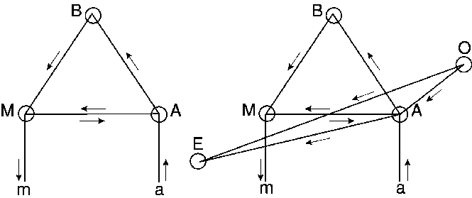
Lichtheim House[Bearbeiten | Quelltext bearbeiten]
-Fragenkatalog_SS18_-_Lichtheim_house.png)
- B = conceptual centre
- M = centre for motor images (Broca)
- A = centre for auditory word images (Wernicke)
- m = motor impulses of speech organs (periphery)
- a = auditory input (nerve, periphery)
- O = optical representation centre for letters
- E = centre for representation and innervation for writing
This is a functional model. Both Lichtheim and Wernicke warned against attempts to localize B, O and E in a single locaction!
I) Transcortical sensory aphasia (A - B)
- Loss of
- understanding of spoken language (auditory words have lost their connections with concepts)
- understanding of written language (the silent reading path O-A-B is also interrupted because involved in A – B)
- Preservation of
- volitional speech (B-M-m intact)
- volitional writing (B-M-A-E intact)
- repeating words (a-A-M-m)
- reading aloud (O-A-M-m)
- writing to dictation (a-A-E)
II) Transcortical motor aphasia (M - B)
- Loss of
- volitional (willful) speech (the concept (B) does not connect to the word movement representation in M)
- volitional writing (B-M-A-E is interrupted)
- Preservation of
- Understanding of spoken language (a-A-B)
- understanding of written language (O-A-B)
- repeating spoken words
- copying words
- writing to dictation (a-A-E)
- reading aloud (O-A-M-m)
Wernicke's proposed classification based on Wernicke-Lichtheim model
- (Cortical) Motor Aphasia (Broca) → (
M) - (Cortical) Sensory Aphasia (Wernicke) → (
A) - Conduction aphasia (arcuate) → (
A-M) - Transcortical motor aphasia → (
M-B) - Transcortical sensory aphasia → (
A-B) - Pure motor aphasia (aphemia, anarthria) → (
m) - Pure verbal (word) deafness → (
a) - Total aphasia (global) → all peri-sylvian cortex
- Amnestic aphasia (anomic, mild) → no location,
B?, ???
This classification has survived til modern times but its shortcomings has led to newer models.
Newer models[Bearbeiten | Quelltext bearbeiten]
American Aphasia Association[Bearbeiten | Quelltext bearbeiten]
-Fragenkatalog_SS18_-_American_Aphasia_Association_types_of_aphasia.png)
Global aphasia[Bearbeiten | Quelltext bearbeiten]
This is the most severe form of aphasia, and is applied to patients who can produce few recognizable words and understand little or no spoken language. Persons with Global Aphasia can neither read nor write. Global aphasia may often be seen immediately after the patient has suffered a stroke and it may rapidly improve if the damage has not been too extensive. However, with greater brain damage, severe and lasting disability may result.
Broca's aphasia ('non-fluent aphasia')[Bearbeiten | Quelltext bearbeiten]
In this form of aphasia, speech output is severely reduced and is limited mainly to short utterances of less than four words. Vocabulary access is limited and the formation of sounds by persons with Broca's aphasia is often laborious and clumsy. The person may understand speech relatively well and be able to read, but be limited in writing. Broca's aphasia is often referred to as a 'non fluent aphasia' because of the halting and effortful quality of speech.
Mixed non-fluent aphasia[Bearbeiten | Quelltext bearbeiten]
This term is applied to patients who have sparse and effortful speech, resembling severe Broca's aphasia. However, unlike persons with Broca's aphasia, they remain limited in their comprehension of speech and do not read or write beyond an elementary level.
Wernicke's aphasia ('fluent aphasia')[Bearbeiten | Quelltext bearbeiten]
In this form of aphasia the ability to grasp the meaning of spoken words is chiefly impaired, while the ease of producing connected speech is not much affected. Therefore Wernicke's aphasia is referred to as a 'fluent aphasia.' However, speech is far from normal. Sentences do not hang together and irrelevant words intrude-sometimes to the point of jargon, in severe cases. Reading and writing are often severely impaired.
Anomic aphasia[Bearbeiten | Quelltext bearbeiten]
This term is applied to persons who are left with a persistent inability to supply the words for the very things they want to talk about-particularly the significant nouns and verbs. As a result their speech, while fluent in grammatical form and output is full of vague circumlocutions and expressions of frustration. They understand speech well, and in most cases, read adequately. Difficulty finding words is as evident in writing as in speech.
Primary progressive aphasia[Bearbeiten | Quelltext bearbeiten]
Primary Progressive Aphasia (PPA) is a neurological syndrome in which language capabilities become slowly and progressively impaired. Unlike other forms of aphasia that result from stroke or brain injury, PPA is caused by neurodegenerative diseases, such as Alzheimer's Disease or Frontotemporal Lobar Degeneration. PPA results from deterioration of brain tissue important for speech and language. Although the first symptoms are problems with speech and language, other problems associated with the underlying disease, such as memory loss, often occur later.
Other varieties[Bearbeiten | Quelltext bearbeiten]
In addition to the foregoing syndromes that are seen repeatedly by speech clinicians, there are many other possible combinations of deficits that do not exactly fit into these categories.Some of the components of a complex aphasia syndrome may also occur in isolation. This may be the case for disorders of reading (alexia) or disorders affecting both reading and writing (alexia and agraphia), following a stroke. Severe impairments of calculation often accompany aphasia, yet in some instances patients retain excellent calculation in spite of the loss of language.
Aphasia, lateralization & music[Bearbeiten | Quelltext bearbeiten]
..” Nonfluent Aphasia: Patients may have trouble putting a three-word sentence together and yet if you ask them to sing an old song, they will sing it fluently,” Patel says. “You wouldn’t know there was anything wrong with them.”
The phenomenon, described in Oliver Sacks’ book, Musicophilia, suggests that there are specialized areas of song in the brain that are not damaged in these patients. Comparing Magnetic Resonance Imaging scans (MRIs) bears this out. “There are distinctive areas, more on the right side of the brain, that are involved with singing “…..“There is a right hemisphere bias in the control of song.”
Dementia: Research has also found evidence that musical memory is retained, even in severe cases of dementia and Alzheimer’s disease.
….“Patients might not know your name,” says Patel, “but if you play familiar songs to them, they might sing along, and know the melody in great detail. If you introduce a wrong note into the song, they will startle, which is evidence they know the details of that melody.”
“Music provides a way to access regions of the brain and reawaken autobiographical memory when language won’t.”
Brain imaging/brain investigation techniques[Bearbeiten | Quelltext bearbeiten]
to be studied for the exam: "Know the basic features about the brain imaging/brain investigation techniques and their advantages / disadvantages (here at least 2), but the very prominent imaging methods (MR-fMRI, DTI, and EEG/ERP/frequency bands) should be learned in more detail."
Methods by type of imaging:[Bearbeiten | Quelltext bearbeiten]
1) Electro-physiological[Bearbeiten | Quelltext bearbeiten]
- EEG (electroencephalography)
- Single unit (single electrodes)
- TMS (transcranial magnetic stimulation)
2) Hemodynamic imaging[Bearbeiten | Quelltext bearbeiten]
- MRI (magnetic resonance imaging)
- fMRI (functional MRI)
- sMRI
- PET (positron emission tomography)
3) Optical imaging[Bearbeiten | Quelltext bearbeiten]
- NIRS (near-infrared spectroscopy)
- DTI (fiber tracking)
- ERP
- TdCS
- MEG (magnetoencephalography)
- ELAN
- Hyperscanning
- synchronization
- P300
Static vs. dynamic[Bearbeiten | Quelltext bearbeiten]
1) static: anatomy/structure
2) dynamic: functional → visualize processes
"Outcome" (trade-off btw. temporal and spatial resolution)[Bearbeiten | Quelltext bearbeiten]
- functional imaging methods require sufficient temporal resolution
- focus on anatomy → emphasis on spatial resolution
Neurolinguistic processing models[Bearbeiten | Quelltext bearbeiten]
to be studied for the exam: "Know at least some neurolinguistic processing models and the names most prominently attached to them."
Lichtheim House[Bearbeiten | Quelltext bearbeiten]
- B = conceptual centre
- M = centre for motor images (Broca)
- A = centre for auditory word images (Wernicke)
- m = motor impulses of speech organs (periphery)
- a = auditory input (nerve, periphery)
- O = optical representation centre for letters
- E = centre for representation and innervation for writing
This is a functional model. Both Lichtheim and Wernicke warned against attempts to localize B, O and E in a single locaction!
Cohort theory[Bearbeiten | Quelltext bearbeiten]
First described by Exner in ~1890, then 90 years later by William Marslen-Wilson
Sounds/phonemes activate neural pathways of possible/predicted words consistent with the phoneme sequence → hearing /s/ activates (neural traces corresponding to) many words beginning with /s/, then hearing /p/ narrows the possibilities (and neural activation pattern) down to words beginning with /sp/ and so on until a single word is 'detected' [similar to autocomplete feature of search engines, text input on smartphones etc.]
Kussmaul's 4 word centres & Charcot's clock diagram[Bearbeiten | Quelltext bearbeiten]
1. stage of “preparation in mind and mood“ ... → thought ... → affective urge which drives to express it.
2. stage of “diction“ , ... → building of internal words together with their syntax, selecting words from memory
3. Articulation and composition of overt words or expressions…
(conceptual preparation – formulation – articulation = still in modern theories of speaking)
4 word centres:
1. centre for motor images,
2. centre for auditory images
3. centre for visual images
4. motor graphic images
Charcot adopted Kussmaul's 4 word centres, developed into 'clock diagram'
IC = ideation centre, CAC = common auditory centre, CVC = common visual centre, CAM = auditory word centre, CVM=visual word centre, CLA = centre for articulate language, CLE = language writing centre
Each word has all 4 components (visual, auditory, motor, motor graphic[?])
→ Theory of psychological types: …Language users are by inheritance and experience, visual, auditive or motor types. One must know a child‘s type for an optimal didactic approach…
Holism (Freud & Hughlings Jackson)[Bearbeiten | Quelltext bearbeiten]
Continuous language region in the cortex with 4 local anchor sites: entrance points of
- acoustic nerve + exit point
- optical nerve + exit point
- speech motor innervation
- handwriting innervation
Speech spans the 4 corners like a tent
Aphasia is always mixed, depending on location, size and severity of lesion; 3 types:
- Pure verbal aphasia [always mixed?!?]
- Agnostic aphasia
- Asymbolic aphasia
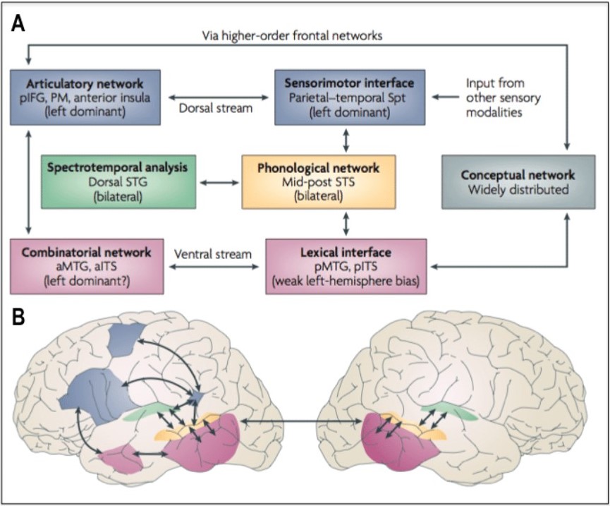
Dual-stream model of language/speech processing (Hickok & Poeppel)[Bearbeiten | Quelltext bearbeiten]
-Fragenkatalog_SS18_-_DualStreamModel.jpg)
Miscellaneous/glossary[Bearbeiten | Quelltext bearbeiten]
- Adrenaline
- amygdala: "Mandelkern"
- angular:
- arbor vitae:
- archicortex:
- astrocytes: Regulate neurotransmitter concentrations; important for blood-brain barrier
- auditory cortex:
- axon:
- basket cells:
- Bereitschaftspotenzial: Neuro-electric potential due to activity in motor cortex and SMA prior to volitional movement
- Betz cells: large motor neurons)
- calcarine:
- caudal: "beak-wards" = posterior
- caudate:
- Cerebellum:
- Cerebrum:
- cingulate:
- claustrum:
- cortex:
- cuneus:
- cytoarchitecture: = "cell architecture", refers to the cellular composition of brain tissue (see also histology); histology (micro-anatomy): science of slicing and staining brain slices (credited to Viennese psychiatrist Theodor Meynert)
- default mode network
- dendrite
- Diencephalon
- dorsal (superior): top side
- dorsolateral prefrontal association cortex
- endorphine (system)
- Epithalamus
- Fissure/Fissura
- folia
- frontal eye field
- Frontal lobe
- genu
- gray matter
- Gyrus
- Gyrus Cinguli/Cingulate Cortex: Attention, impulse, drive, motoric processing, emotions, mimicry, gesture…
- HF Hippocampal Formation
- Hypothalamus
- Inferior
- limbic system
- marginal
- Medial Gyri: Below the surface gyri in the cortex → only visible in cross-section visualizations; esp. Gyrus Cinguli in Cingulate Cortex
- Medulla oblongata
- Mesencephalon
- microcytes (microglia)
- myelin
- myelin sheath
- neocortex
- neuron
- Noradrenaline
- Occipital lobe
- oligodendrocytes (oligo = few, dendron = tree) enwrap axons → myelin sheath
- PAG (peri-aqueductal gray) part of endorphine system
- parietal
- Parietal lobe
- peri-sylvian language network
- phrenology: deriving mental faculties and character traits from the shape of someone's skull
- planum temporale
- pons
- posterior parietal assocation cortex
- posterior parietal cortex
- prefrontal cortex
- Prefrontal lobe
- premotor cortex
- primary motor cortex
- primary sematosensory cortex
- process (of a neuron ) appendix (unipolar = 1 "process" (appendix), bipolar = 2 appendices: 1 axon, 1 dendrite, multipolar = ...
- pSTG posterior part of Superior Temporal Gyrus
- Purkinje cells
- Purkinje cells (large cells in cerebellum)
- Pyramidal cells
- ramus
- reward system
- rostral (frontal, anterior)
- rostrum
- Serotonine
- SMA (supplementary motor area/cortex)
- soma
- somatosensory cortex
- splenium
- substantia nigra
- Subthalamus
- Sulcus
- Superior
- supramarginal
- synapsis
- Temporal lobe
- Thalamus
- Thalamus
- ventral (inferior): bottom side
- ventricle: "cavity"
- visual cortex
- white matter